Fluke Biomedical 07-661-7662 User Manual
Page 9
Introduction
Image Intensifier
1
1-5
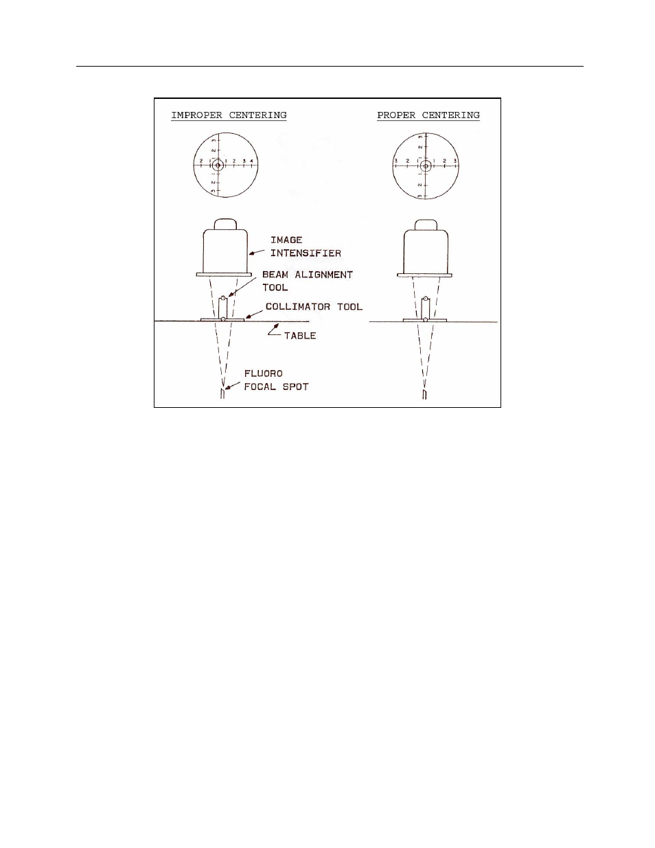
Figure 1-6. Placement of Collimator and Beam Alignment Test Tools for Checking Fluoro Units
When this occurs, the alignment of the center of the x-ray tube to the image intensifier can be checked by
noting if the image of the Collimator Tool is symmetrical, as seen on the TV monitor or mirror. While
observing the image of the centimeter scales of the Collimator Tool, record the distance to the edge of the
viewing field for each of the four axes of the tool. If the x-ray field is centered, these distances should be
the same (Figure 1-6). If the centering is off by more than 3% of the SID, it should be corrected by
qualified service personnel.
Tape a large cardboard cassette, containing a fresh film or a "Ready-Pac", to the input face of the image
intensifier. Open the fluoro collimator shutters as far as possible, and expose the cassette in the fluoro
mode for about 15 seconds at 60 kVp and 1-2 mA. The spot film device is not used for this test exposure.
The processed film will show the image of the Collimator Tool. The extent of the actual field size at the
tabletop will be shown as the distance on the Collimator Tool scale that is imaged for each axis of the
tool. The difference between the viewed field size and the actual field shown on the film is a collimation
error. This test is illustrated in Figure 1-7.