Fluke Biomedical 6000-529 User Manual
Page 10
Victoreen 6000-529
Operators Manual
2-2
with enough aluminum to give a reading just below one-half the unattenuated reading and that made with
aluminum to give a reading just greater than one-half the unattenuated reading.
When making beam quality measurements at mammographic energies, it is recommended that you use
99% pure aluminum. Alloy 1100 aluminum has been used, however studies show that an error as great
as 7.5% may result.
To perform a beam quality test, follow these steps:
1. Raise the compression paddle to its highest position. Mount the 6000-529 ionization chamber on a
ring stand so there is approximately 5 cm of space between the bottom of the chamber and table.
The chamber should be centered in the beam laterally, and approximately 4 cm from the chest wall.
2. Collimate the beam, using the light field, so that the entire chamber is included in the beam. The
field should be approximately 6 cm x 6 cm. If necessary, relocate the chamber such that it is
centered in the field.
3. Set the kVp selector at a kVp setting that is frequently used for making mammograms. Set manual
timing, and set the mAs to provide an exposure reading of at least 500 mR. Refer to the instruction
manual for the electrometer or other instrument used to measure charge generated in the 6000-529
chamber.
4. Make an exposure. Note the reading and label it X
0
. If you are using an electrometer that reads in
nC or some other instrument that would normally require the application of a correction factor, you
may note the raw reading without corrections. This is so because all subsequent readings are
normalized to X
0
.
5. Place a sheet of aluminum 0.2 mm thick on the compression paddle. Using the collimator light, be
sure the entire ionization chamber is in the shadow of the aluminum sheet. Make an exposure.
Record the reading and label it X
1
; also record the thickness of aluminum used to make the
exposure. Label it t
1
.
6. Place an additional 0.01 mm of aluminum on top of the aluminum absorber(s) already in place.
Make an exposure. Record the reading, labeling it with sequential indices. Also, record the total
thickness of aluminum used in making the measurement, labeling it as t
N
where N is the total
number of filtered exposures taken so far. If X
N
is less than one half of X
0
proceed to step 7,
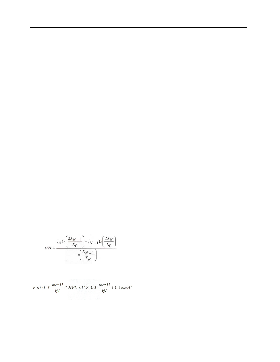
otherwise, repeat step 6.
7. It is now assumed that you have compiled a list of data pairs, labeled "t
i
" and "X
i
"- If N is the total
number of filtered exposures, then the half-value layer may then be calculated using the following
formula:
It is recommended that the HVL be in the following range:
If your calculated HVL is lower than this range, you may be in violation of Federal or State regulation. For
more information, see American College of Radiology Medical Physicist's Manual.