Fluke Biomedical 07-611 User Manual
Page 13

Operation
X-Ray Tube Focal Spot Measurements
2
2-7
[
]
[
]
[
]
2.1.7 Procedure - Slit Measurement of Focal Spots
1. Replace the test stand alignment device with the slit assembly, parallel to the anode-cathode axis.
2. Place a direct exposure x-ray film or cassette under the base for over-table x-ray tubes or on top of
the stand for under-table tubes.
3. Using the x-ray tube rating chart, select a technique of about 75 kVp and one-half the maximum
rated mA at 0.1 sec exposure for the appropriate focal spot size.
4. Select the exposure time to obtain about 300 mAs for the direct exposure film or 30 mAs for the
cassette at a 36-inch (90 cm) source-to-image distance (film density should be between 0.8 and 1.2
above the base-plus-fog of the film).
5. Align the slit assembly parallel to the anode-cathode axis to make the focal width measurement.
6. Expose the cassette or film at the selected technique factors (steps, 3 and 4, above).
7. Move the cassette a few inches (to prevent double exposure).
8. Rotate the slit assembly 90° to measure the focal length.
9. Expose the cassette or film at the selected technique factor.
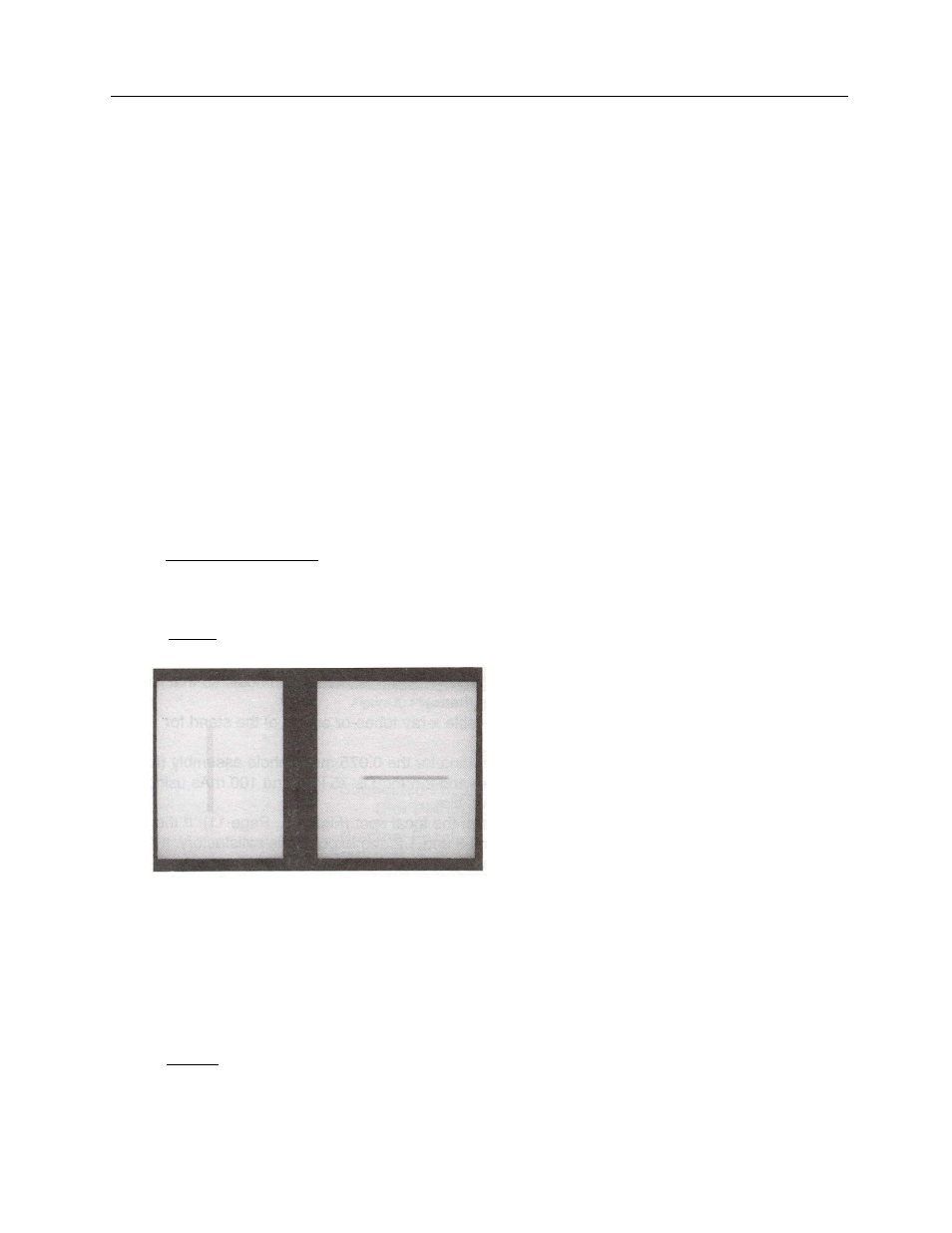
10. Process and view the slit images (Figure 2-9).
11. Measure the center-to-center distance between the localization holes on the radiograph using the
ruler.
12. Divide image localization hole separation by the small adapter ring localization hole distance (40
mm). Calculate the magnification using the formula below:
image hole separation
40
mm
- 1 = Magnification
For example, assume the image hole separation was measured as 90 mm
90 mm
40 mm
- 1 = 1.25
Figure 2-9. Slit Image Parallel and Perpendicular to the Anode-Cathode Axis
13. Measure across the middle of each slit image using the magnifier lens (with a built-in graticule). The
measurement across the band parallel to the anode-cathode axis is related to the width of the focal
spot. The measurement of the band perpendicular to the anode-cathode axis is related to the length
of the focal spot.
14. To determine focal spot size, divide the measured width and length by the magnification factor. For
example, if the measured width of the slit image measured 1.8 mm then
1.8 mm
1.25
= 1.44 mm (width of focal spot)