Fluke Biomedical 4000M+ User Manual
Page 23
3
Theory of Operation
Detector Positioning
3-5
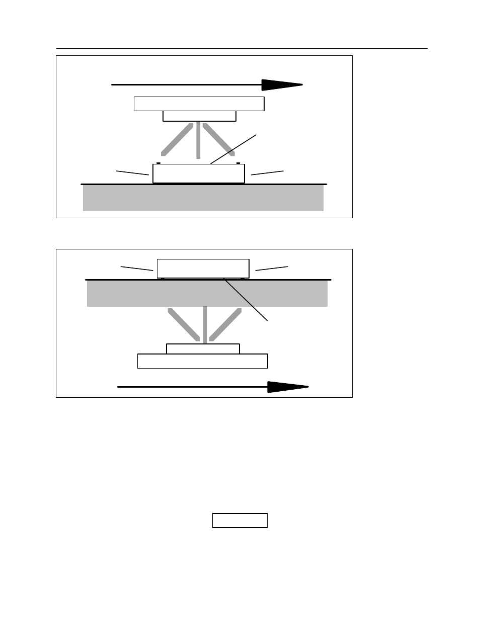
X-RAY TUBE
TUBE AXIS
MODEL 4000+
TOP PANEL
FRONT OR REAR PANEL
FRONT OR REAR PANEL
Figure 3-1.
Detector Positioning (Radiographic Measurements)
X-RAY TUBE
TUBE AXIS
MODEL 4000+
TOP PANEL
FRONT OR REAR PANEL
FRONT OR REAR PANEL
Figure 3-2.
Detector Positioning (Fluoroscopic Measurements)
3. Use the collimator light to collimate the beam to a rectangular area the same dimensions as the top
of the detector box. The collimated beam should be approximately 22 square centimeters (8-1/2
by 9 inches).
4. Move the detector so that the black disk is centered in the collimated beam.
Fluoroscopic Measurements
In the Fluoroscopic Mode, the Model 4000M+ must usually be turned upside down. The black plastic disk
must face the x-ray tube, which is normally located under the table.
NOTE
When the unit is upside down in the Fluoroscopic
Mode, the Display automatically inverts so it can be
read in that position.