Arrhythmia functions, Atrial fibrillation – Fluke Biomedical MPS450 User Manual
Page 42
MPS450
Operators Manual
2-10
Table 2-8. Numeric Codes for Pacemaker-Width Settings
Pacemaker-Width Setting
Numeric Code
0.1 ms
243
0.5 ms
244
1.0 ms
245
1.5 ms
246
2.0 ms
247
Arrhythmia Functions
Departures from the normal height, shape, or length (of time) of the PQRST-waveform
patterns suggest specific illnesses, making the ECG very valuable when used in
conjunction with other diagnostic tests. ECG patterns that divulge disturbances in the
blood supply to the heart muscle or abnormalities in the heartbeat (arrhythmias) may be
associated with coronary artery disease.
The MPS450 simulates a wide array of arrhythmias, representing heartbeats that are too
slow, too fast, or totally erratic; that have beats with abnormal timing, spacing, or
waveform shapes; or that combine abnormal and normal beats in varying proportions.
Atrial Fibrillation
A rapid, irregular atrial signal, coarse or fine, with no real P waves; an irregular
ventricular rate.
Coarse and fine atrial fibrillation occurs when the electrical signals in the atria are
chaotic, and multiple, ectopic pacemakers are firing erratically. Some impulses may
conduct through to the AV node to stimulate the ventricles, causing a quite-irregular and
often-rapid ventricular rate. On the ECG there is an absence of P waves, with an irregular
R-R interval. Atrial-fibrillation waveforms are irregularly shaped and usually rounded.
The amplitude of the atrial signal is higher for coarse, and lower for fine, fibrillation.
The MPS450 simulates irregular atrial waveforms; the amplitude of the atrial signal is
higher for coarse, and lower for fine, fibrillation.
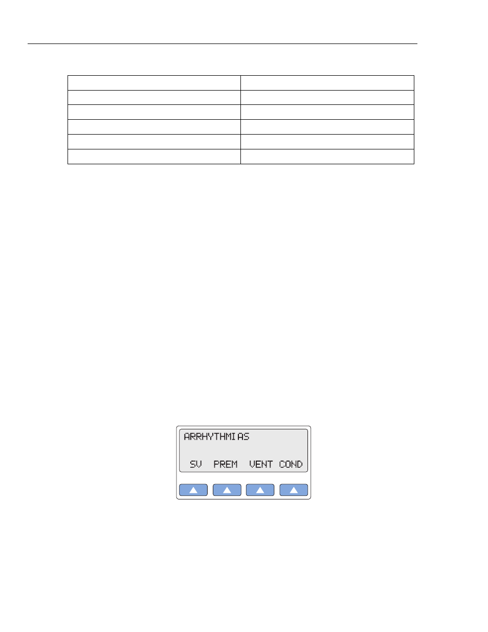
Action in the Menu-Control Mode
1. Press the top-menu key labeled ARRHY to display the following LCD screen:
gje015.eps
2. Select SV to display the following LCD screen: